
What is spatial (3D/4D) ultrasound good for?
Modern ultrasound technology allows us to immediately create a three-dimensional image. (If we can only create a still image, we call it 3D, if we see a continuously moving image, we call it 4D). This significantly simplifies the examination, not to mention the fact that it is sufficient to take a single good spatial image, which can be cleaned afterwards, rotated in space, and certain important parts highlighted. Consider that in early pregnancy we use a vaginal probe, so the examination in two dimensions takes much longer than creating a good three-dimensional image and then shaping the image afterwards (that is, without burdening the expectant mother).
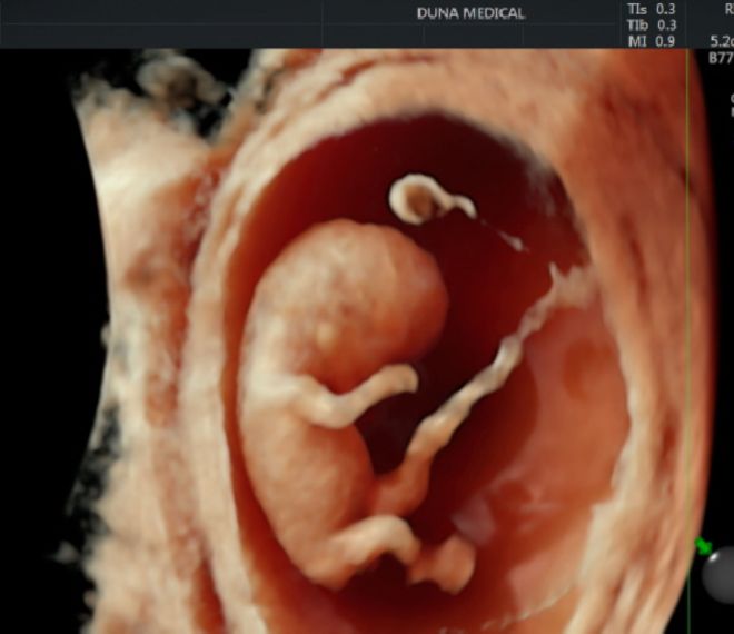
Let's see an example! And a riddle: how old is the fetus in the pictures? (The answer can be found at the end of the pictures.)
This is the basic image:

After post-processing, we can see this from above:

This is how we see the fetus from below:

From the eye like this:

From the other side:

And twisted a little more:

The answer to the riddle is: the embryo shown in the pictures is 9 weeks old!
The pictures were taken on the GE Voluson E10 device of the Duna Medical Center.
If you would like to book an appointment for a gynecological examination, you can do so via our telephone customer service at +36 1 790 7070 or online !
Get to know our gynecologist who performed the above examination: