
 Radiology is the umbrella term for diagnostic imaging tests. Imaging diagnostics can be used to image any area or organ of the body in great detail. Most radiological examinations, with the exception of ultrasound diagnostics, are performed using X-rays.
Duna Medical Center's radiology department is equipped with state-of-the-art, high-tech equipment. In addition to the quality of the equipment, the expertise and experience of the radiologist and radiology assistants have a decisive influence on the evaluation of the examinations, which is why our patients are welcomed by highly qualified and experienced professionals.
Radiology is the umbrella term for diagnostic imaging tests. Imaging diagnostics can be used to image any area or organ of the body in great detail. Most radiological examinations, with the exception of ultrasound diagnostics, are performed using X-rays.
Duna Medical Center's radiology department is equipped with state-of-the-art, high-tech equipment. In addition to the quality of the equipment, the expertise and experience of the radiologist and radiology assistants have a decisive influence on the evaluation of the examinations, which is why our patients are welcomed by highly qualified and experienced professionals.
CT scans
CT scans use an X-ray beam to take slice images of specific regions of the body. The series of images are evaluated by a radiologist using various software, and his findings, diagnosis and any further action to be taken are described in a text report. The hundreds - sometimes thousands - of images taken during the examination can be used to diagnose lesions that cannot be detected by other imaging diagnostic methods. When we need a more precise picture of the function of an organ or a lesion in an organ, we may need to use intravenous contrast.
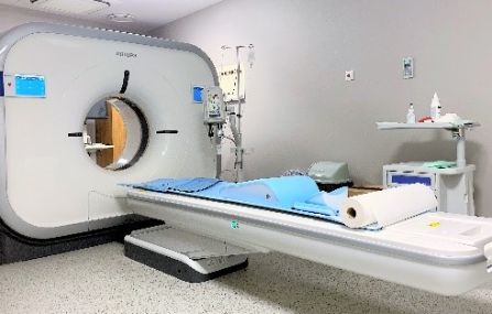
At the Duna Medical Center, a 128-slice Philips Incisive Pro CT scanner is used to accurately identify abnormal lesions. The equipment has a number of special software features that are not available in one place in our country. This state-of-the-art, top-of-the-range device produces images with extremely high spatial resolution thanks to a minimum slice thickness of 0.625 millimetres. The very fast rotation speed of 0.35 seconds results in images that do not shift. The high-sensitivity NanoPanel Elite detector system ensures the lowest possible radiance. Most of our CT scans are measured in 1-3 breaths.
Thanks to the software of the device, excellent images can be obtained even with embedded metals, such as hip prostheses. Radiologists are aided by built-in artificial intelligence to detect lung and colon tumours. The device is suitable for low-dose CT lung screening. Other special capabilities include accurate size tracking and comparison software for lung lesions, 3-dimensional imaging of the vascular system, and oncology tracking software to measure changes in tumours as a result of treatment.
An important feature of the device is that it can be used to examine patients with a high body weight, up to 205 kilograms.
The total time for CT scans, after any preparation, is usually 15-60 minutes. After a contrast agent scan, 20 minutes should be spent in the waiting room to identify and manage any side effects caused by the contrast agent.
CT scan procedure: the scan is performed in the supine position and is painless. For chest and abdomen scans, the arms should be placed above the head so as not to interfere with the imaging. Removable metal objects such as necklaces, earrings and removable dentures should be removed as they also interfere with the imaging. Avoid eating for 6 hours before the scan, but drink plenty of water. Regular medication should be taken with water before the examination.
Do not move around during the scan. A series of measurements should be taken in 5-30 seconds. For chest and abdominal examinations, breathing must be held for this time, as instructed by the operator via microphone. In some cases, for abdominal and pelvic examinations, 1-1.5 litres of water or diluted contrast medium must be drunk approximately 1-1.5 hours before the examination. This is necessary to separate the intestines from their surroundings and to allow the intestinal wall to be examined. For examination of the rectum and colon, it may rarely be necessary to fill the rectum or colon - enema. For cardiac examinations it may be necessary to administer a preparatory drug. For these special tests, the total time of the test including preparation may be up to 1.5 hours.
For certain examinations, the administration of contrast material is prescribed. This contrast material is an iodine-containing compound suitable for the imaging of blood vessels and the examination of blood circulation in organs and tissues. It is most often introduced into the body through a vein in the arm. Among the regularly taken medications, the administration of iodine-containing contrast material should be avoided with certain medications used for diabetes. These antidiabetic drugs cannot be taken for 2 days before and after the contrast-enhanced examination. The method of suspending the medication should be discussed with the treating physician.
With normal kidney function, the contrast material does not harm the kidneys, but it is important to consume plenty of fluids on the days before and after the administration of contrast material!
As with any drug, hypersensitivity to contrast material can occur rarely. The hypersensitivity is not to the iodine itself but to the combination of iodine and the molecule it is bonded to, that is, to the compound.
For abdominal and pelvic CT scans, drinking a different type of contrast material is also necessary to better assess and differentiate the intestines. This contrast material can cause diarrhea.
CT scans during pregnancy are only conducted in special cases or if there's a suspicion of a serious illness, as X-ray radiation can harm the fetus. CT scans can be safely performed during breastfeeding. If a venous contrast material examination is needed, do not breastfeed for two days following the administration of the contrast material; instead, express the milk but do not feed it to your child.
A referral from a physician is required for CT examinations. This referral must include the reason for the examination, the patient’s symptoms, relevant medical history, preliminary diagnosis, as well as the exact region to be examined and the type of scan requested.
To ensure accurate planning and high-quality imaging and reporting, it is essential that this information is provided by the referring doctor – such as the patient's general practitioner or specialist. Without these details, the examination cannot be performed.
It is the patient’s responsibility and in their best interest to ensure all necessary documentation is obtained and provided.
The evaluation of the results is completed within a maximum of 3 working days following the examination. The results can be accessed electronically via the EESZT (Electronic Health Service Space) through the Client Portal (Ügyfélkapu), collected in person at a pre-arranged time or via an authorized representative, or sent by email. The preferred method of receiving the results should be arranged with the assistant performing the examination and the reception staff.
The imaging records can be provided exclusively on a CD upon the patient’s prior request.
Please read and fill out our patient information form, and bring it with you to the examination to ensure a smooth and quick administrative process.

If you require a medical report in a foreign language (English, German) please indicate this to our staff during your visit, as preparing these involves an additional cost. The documents are prepared within 3-10 working days.
-
CT - Only contrast examination per regionFrom 55 000 Ft
-
CT Abdominal - pelvic examination nativelyFrom 62 000 Ft
-
CT Abdominal pelvic examination, natively and with contrast materialFrom 106 000 Ft
-
CT Abdominal-pelvic examination natively and supplemented with urography with contrast materialFrom 114 000 Ft
-
CT Angiography chest or abdomen reconstructed - by regionFrom 106 000 Ft
-
CT Chest-abdominal-pelvis examination nativelyFrom 92 000 Ft
-
CT Chest-abdominal-pelvis examination natively and with contrast materialFrom 128 000 Ft
-
CT examination native, by region (cranium, facial skull, inner ear, cervical soft tissue, orbit, cervical spine, dorsal spine, lumbar spine, hip, limb, chest)From 42 000 Ft
-
CT examination per native + contrast region (cranium, facial skull, cervical soft tissue, chest)From 84 000 Ft
-
CT lung screening - native examination with reduced radiation exposureFrom 35 000 Ft
-
CT neck-chest-abdomen-pelvis examination nativelyFrom 106 000 Ft
-
CT neck-chest-abdomen-pelvis examination natively and with contrast materialFrom 128 000 Ft
-
CT scan - Angiography of lower limb reconstructed (both limbs are examined)From 256 000 Ft
-
CT skull + carotid angioFrom 128 000 Ft