
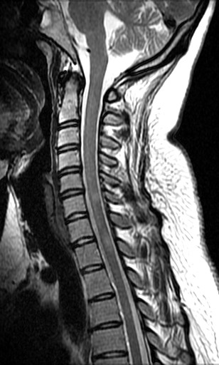
Magnetic resonance imaging (MR, MRI) scan
The aim of the study
MR scans can provide high-resolution, information-rich images of many organs and organ systems, allowing a wide range of diseases and conditions to be investigated. Among these, the nervous system (brain, spinal cord), joints and extremities, male and female pelvis, abdominal organs (liver, bile ducts, spleen, kidneys, adrenal glands, pancreas, etc.), breast and certain blood vessels, heart are of great importance.
 |
 |
The scan can be used to visualise various inflammatory and tumour lesions, blood supply disorders, injuries, developmental disorders and vascular blockages. MR is a non-invasive method, but MR imaging can also be used to perform special tissue sampling (biopsy)
MRI scanning is one of the most modern imaging diagnostic techniques. It uses a computer to take images of a specific part of the body in a strong magnetic field in different planes. The procedure does not involve any radiation exposure and, as far as we know today, has no proven adverse health effects. However, it can be a risk for all patients who have had a surgical implantation of a magnetised metal containing metal (heart valve, vascular stimulator, neurostimulator, internal hearing aid, joint prosthesis, drug injection device) or who have had an accidental ingestion of a metal foreign body (metal shrapnel, metal fragments, bullet). Devices and objects containing magnetisable material (iron, steel) may shift or heat up in the magnetic field, causing serious, life-threatening injuries or rendering the examination useless. Be sure to inform the examining operator or radiologist of the presence of this/these. It is the radiologist's responsibility to assess this, so please bring your final report and related documents with you. Any metal objects worn on the body (hearing aid, watch, chain, metal clasp, body jewellery, hair clip, wave clip, etc.) must be removed and pockets emptied (metal coins, keys, magnetic card, pen, etc.) must be placed in the changing room in the changing room. If you have metallic cosmetics (mascara, powder, magnetic mascara) on your face or are wearing a wig, be sure to remove them.
Medicines that you need to take regularly can be taken with a little water on the day of the test. You should not eat for 4-6 hours before the test, but you can drink if possible, because of the possibility of contrast media being given.
Information before the test
- Before the test, all medicines can be taken as usual and fluids can be consumed safely. It is not necessary to come on an empty stomach, but you should not eat for two hours before the test because of the possible administration of contrast media.
- Before the test, the patient will receive a written information leaflet and will be asked to answer the questions on it in writing. The patient must indicate if he/she has a metallic device in his/her body (e.g. pacemaker, prosthetic joint, heart valve, middle ear, metallic device from an accidental operation, implanted pump, etc.) or if he/she has been involved in an accident where metal fragments, metal shrapnel or bullets may have entered his/her body or eyes. The date of implantation is also important information. In addition to the above information, the patient should also indicate if he/she has any kidney dysfunction, which is important for contrast administration.
- In pregnancy, the test is only performed in very justified cases. Although there is no evidence of adverse effects on the foetus, some effects are not yet clear, so it is better to be cautious.
- When the patient arrives for the scan, remove all metallic materials from the body (e.g. jewellery, watch, hair clip, brooch, necklace, belt, braces, glasses, zipper, clasp, removable prosthesis, hearing aid, mobile phone, etc.) - all these will reduce the quality of the scan and may be displaced in the magnetic field. Strong magnetic fields can damage all sensitive items (credit cards, debit cards, library cards), so be sure to leave them in a lockable changing room. For some examinations and depending on the patient's clothing, a change of clothes is required, for which disposable clothing is provided.
- If you have tattoos or are wearing medicated plasters, please also inform the examining assistant as these may contain small amounts of metallic material which may heat up during the examination.
- You will also receive a consent form with the information leaflet, and you must answer the questions on this form (whether you agree to the test, whether you agree to the contrast medium) in writing with a yes or no.
- The test takes 20-60 minutes. It depends a lot on which part of the body is being examined and what is wrong with it. The test is done in the lying position and is painless. The only inconvenience is that the test is performed in a relatively confined space, which can cause a feeling of confinement (claustrophobia) in some people. The LIV DMC is roomier and shorter than the standard one, so it can be used for most patients with known claustrophobia - please let us know in advance when you book!
- If you experience any anxiety or other problems during the examination, please let the examining operator know and they will monitor you throughout the examination.
- The machine will make a tapping, buzzing sound while it is operating, which is when the images are taken. Patients must lie still in the position set by the operator during the scan, otherwise the images will be unreadable to the doctor due to the disturbing artificial products caused by the movement.
In many cases, a contrast agent injected into a vein is needed to better assess anatomical images - this allows better differentiation between organs, more accurate detection of abnormal lesions and visualisation of blood vessels.
There are four features of an MRI scan that are essential to know before the scan is performed
- The confined space: some patients have a feeling of confinement, which in most cases disappears in the presence of a relative. It should be remembered that in the examination room, the patient is not confined, but is in close contact with the staff (audio, video).
- Noise: the device may emit a loud buzzing, humming or knocking sound during the measurements. This noise may last for 3-10 minutes. The noise is caused by the operation of the magnets. This is when the actual test takes place, when you have to lie still.
- Heating: the temperature inside the magnet is 22-25 degrees Celsius, the test room is air-conditioned.
- The scan is relatively lengthy: probably the most difficult part of the MR scan is lying still. Any movement of the patient greatly reduces the quality of the resulting images.
After the examination is completed, the assistant removes the coils from your body and informs you that you may get up and get dressed. At this point, the imaging process may not yet be fully finished, as additional images and post-processing may be required based on the acquired data.
Following this, the examination is evaluated by a radiology specialist, which requires additional time. The assistants performing the examination can provide information on when your report is expected to be ready. Once completed, the report can be collected from the patient administration desk.
The imaging files can be provided exclusively on a CD upon prior request from the patient.
What machine do we use for our MR scans?
- 70 cm, wider than usual tube diameter (most models are 60 cm) so the patient will have a more spacious experience during the examination.
- Shorter tube length - the machine is equipped with a magnet only 137cm long - which allows patients to see over the top of the tube during the following examinations (very important for claustrophobic patients):
- Lower extremity joints (ankle, leg, knee, hip)
- Pelvis (prostate)
- Examination table load capacity 250 kg - allows for more serious patients!
- Extensive software back-up (shorter examination time, better resolution images)
- Manufacturer dedicated special coils for very detailed examination of many organ systems.
General information on radiological examinations
- Medical referral: private MR scans do not require an official referral. In the case of a medical referral, in order to personalise the examination and answer the clinician's questions, we recommend that your doctor describes in any form what medical conditions you have had, what treatments (surgery, medication) you have had, and the guiding diagnosis or clinical question you are expecting the MR examination to answer.
- Documents: identity card
- Medical history: it is important that you bring your previous final reports, medical records and, if available, the findings and images of your previous radiological examinations (on film or CD). Medical records are a great help in making an accurate diagnosis and in determining the changes in your condition (improvement, deterioration). If we can compare the abnormalities found in the examination with the previous condition, we can give a much more accurate opinion of your body.
Pregnancy:
If there is any possibility that you may be pregnant, please inform us at the time of booking! This must be clarified before certain radiological examinations.
Allergies:
In our department, contrast agents and local anesthetics are most commonly used. Please inform us prior to the examination if you have any known sensitivity or allergy to medications (including contrast agents).
Please also inform us in advance if you have any kidney disease, thyroid disorder, or diabetes. In the case of diabetes, it is particularly important to know the names of the medications you are taking, as these may significantly influence the outcome of the examination.
Clothing and (body) jewelry:
Please prepare in a way that allows for easy changing of clothes before and after the examination.
For certain examinations, it may be necessary to remove clothing covering the examined body part, as well as jewelry (earrings, necklaces, etc.), body piercings, and removable dental prostheses, as these may interfere with image quality. We kindly ask you to prepare accordingly to minimize any inconvenience.
Consent form:
For certain procedures, completing and signing a consent form is required. Prior to the examination, the attending physician or assistant will be happy to answer any questions you may have.
Order of examinations:
In rare cases, we may need to perform urgent (acute) examinations for patients in serious condition (e.g. stroke). Therefore, exceptionally, it may occur that we are unable to start your examination exactly at the scheduled appointment time, and you may be required to wait. We kindly ask for your understanding.
Report issuance:
During a radiological examination, several thousand images may be taken within a few minutes. The evaluation and precise comparison with previous findings require thorough and careful work, and may also involve consultation with your referring physician by phone or in person. This process requires time for the reporting radiologist.
Imaging files can be provided exclusively on a CD upon prior request from the patient.
About contrast media used in radiological examinations
Contrast media are often used in radiological examinations. Contrast agents are used to make organs and tissues visible, to identify them better and to diagnose diseases and pathological conditions more easily, with greater certainty and detail. Their name refers to the fact that normal organs or pathological abnormalities will be more distinct, with a greater contrast difference in the image compared to their surroundings, than without them. The administration of contrast agents can often better distinguish between diseases and normal conditions, and some diseases or pathological conditions may behave in a characteristic way when contrast agents are administered.
Contrast media are used for certain X-ray examinations (including oesophageal and gastric X-rays, colonoscopy (irrigoscopy), urography ("kidney staining"), various body cavity filling examinations) and very often for CT and MR scans. Invasive, catheter-based vascular staining (angiography) may be performed using contrast only. Contrast may also be required for blood tests and there are contrast agents for ultrasound examinations, but these are not yet widely used.
The principle of enhancing contrast
The contrast agents used in X-ray diagnostics (as well as CT and angiography) predominantly absorb X-ray radiation with increased absorption in tissue, which is how the organs and lesions they visualise become visible. These are called positive contrast agents. The contrast agents ingested, passed through the rectum or otherwise into body cavities fill the inside of organs and lesions, displaying their inner surfaces and making them visible. In some cases, so-called negative contrast media (less absorbent of X-rays than tissue) are used, most commonly in the gastrointestinal tract, which may simply be air or carbon dioxide released from a special powder to help visualise the gastrointestinal mucosa more accurately.
Contrast agents administered into the blood vessels (thus intravenously) are carried by the bloodstream to many points in the body where, depending on the properties of the organs, they are excreted, appear in the organ and their pathological lesions can be more accurately and more reliably visualised. This is usually expressed in the findings as the accumulation of contrast material in the organ or lesion, and important conclusions can be drawn from this fact and from the speed and extent of the accumulation.
Contrast media can be classified according to their use or composition.
Contrast media commonly used in the gastrointestinal system are those containing barium sulphate, a white liquid made up of powder and water.
X-ray, CT, angiography contrast media contain iodine bound to various compounds.
In some cases (emergency and post-operative conditions, ingested foreign bodies, etc.), iodine-containing contrast media are also used for the gastrointestinal system, but are not normally absorbed from the intestinal tract.
The contrast agents used for MR scans are special, most often containing compounds of the element gadolinium, which influence the behaviour of organs and lesions during MR imaging by providing them with higher contrast.
Side effects of contrast media (Intravenous contrast media):
CT, vascular staining, urography contrast agents:
Iodine-containing contrast agents given into the bloodstream are rare, but several complications and side effects are possible.
Adverse reactions may include allergic reactions, minor skin redness, hives, itching, subcutaneous oedema, and rarely more severe symptoms such as bronchospasm, bronchospasm, pulmonary oedema. These most often develop soon after contrast administration, but in some cases mild allergic reactions may occur later, even a few days after contrast administration. The way they develop is not exactly the same as allergies of other origins (e.g. drug, pollen, etc.), as they do not require a prior sensitising encounter with the contrast agent and may occur for the first time.
Non-allergic side effects of contrast media include headache, fever, abdominal discomfort, hypertension and very rarely, in the case of iodinated contrast media, swelling of the salivary glands. Very rare are neurological complications such as seizures, transient sensory disturbances.
Deterioration of kidney function can occur almost exclusively in patients with already reduced kidney function. Patients with hyperthyroidism may experience a severe, even life-threatening flare-up of symptoms.
In the case of beta-blocker-type antihypertensive and cardiac rhythm-controlling drugs, allergic nausea with hypotension may occur in rare cases.
In very rare cases, contrast agents administered locally into the blood vessels may cause inflammation of the vessel wall, venous thrombosis. Contrast agents injected into the arteries may cause local side effects, see the leaflet on angiography for more information.
Allergic complications of contrast agents given during MRI scans are similar to those of iodinated contrast agents, but are also much less common. In patients with severe renal dysfunction, some gadolinium-containing contrast agents may develop a late complication of nephrogenic systemic fibrosis (NSF) with connective tissue degeneration of the skin and other organs.
Declaration of consent
Due to the potential risks, it is a legal requirement that the patient must sign a written informed consent form, i.e. sign that they agree to the administration of contrast media, after reading the information leaflet and the patient information leaflet before the tests. In the event that he or she is unable to do so due to his or her condition, a close relative or legal representative, as defined by law, may sign. A declaration of consent is not required if the patient is in imminent danger of death or is in a serious condition and obtaining a declaration would result in a delay which could lead to serious or permanent damage to the patient's health.
Patients should avoid solid food for at least 4 hours before the tests to reduce the risk of nausea and vomiting. It is also recommended to stop drinking fluids 2 hours before the test, but it is not necessary or advisable to be thirsty for longer.
The day after the test, it is also recommended to drink plenty of fluids to protect kidney function, or if this is not possible for some reason, intravenous fluid replacement.
To reduce the possibility of other side effects and complications, it is necessary to be aware of the other diseases and medications described above, and the patient will be questioned about these by the assistants before the test, consulting the examining doctor if necessary. In case of doubt, the risks will also be discussed with the referring doctor.
As mentioned above, complications of an allergic nature are unpredictable, but the investigators and doctors are prepared for them if they occur and will intervene as soon as possible to treat them.
No intravenous contrast agent should be given to patients who have already been shown to have adverse reactions to the contrast agent. Since contrast agent allergy is not to iodine but to the whole molecule, the administration of another type of contrast agent is not contraindicated but requires great caution.
The iodine used for surgical lavage is not the same as contrast agents, so an allergy to iodine-based lavage products does not mean a contrast agent allergy, which does not exclude the use of contrast agents.
In patients with proven hyperthyroidism, iodinated contrast media should not be given because of the serious consequences of thyroid metabolism upset. In the case of persistently treated and well controlled hyperthyroidism, contrast media can usually be administered at the discretion of the endocrinologist treating the patient.
In patients with renal insufficiency, severe renal dysfunction, intravenous iodine contrast should not be given, except in very severe and emergency conditions where the correct diagnosis of the pathological condition is more important than the risk associated with contrast. In these cases, too, the adverse effects of contrast administration are minimised by providing plenty of fluids before and after the examination and by preparation with medication.
Important information for people with diabetes on the pill
For diabetics, certain tablets should be stopped 48 hours before the test and can be started again two days after the test, depending on the results of the blood test (kidney function). If necessary, consult your doctor. For detailed information, please call us on +36 1 790 7070 or send an e-mail to info@dunamedical.com.
Please read and fill out our patient information form, and bring it with you to the examination to ensure a smooth and quick administrative process.

If you require a medical report in a foreign language (English, German) please indicate this to our staff during your visit, as preparing these involves an additional cost. The documents are prepared within 3-10 working days.
-
Abdomen and pelvis non-contrast MR examinationFrom 98 000 Ft
-
Abdominal MRI and MRCP non-contrast+with contrastFrom 108 000 Ft
-
Breast MRI examination natively - to evaluate the implantFrom 85 000 Ft
-
Breast MRI examination natively and with contrast - to evaluate the glandular tissueFrom 105 000 Ft
-
Breast MRI examination natively and with contrast - to evaluate the glandular tissue and implant togetherFrom 115 000 Ft
-
Liver-specific MR scanFrom 148 000 Ft
-
MR - English language reportFrom 31 000 Ft
-
MR abdominal-pelvic exam. non-contrast + contrastFrom 168 000 Ft
-
MR angiography by region - non-contrastFrom 58 000 Ft
-
MR Brain non-contrast + contrast + angiographyFrom 108 000 Ft
-
MR contrast examination only per regionFrom 68 000 Ft
-
MR Cranial non-contrast + angiographyFrom 75 000 Ft
-
MR whole spine non-contrastFrom 128 000 Ft
-
MR Whole spine non-contrast + contrastFrom 168 000 Ft
-
MRI scan non-contrast + contrast per regionFrom 98 000 Ft
-
MRI scan non-contrast, by regionFrom 53 000 Ft
-
Multiparametric prostate MRFrom 98 000 Ft