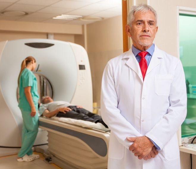
X-ray examination - general information

X-rays can be taken:
- mainly about the bones that make up the body's skeletal system (due to injury, degenerative deviation, suspected fracture, bone change, exclusion of other pathological processes, developmental disorder)
- about joints (in case of bruises, chronic pain, etc.)
- about complaints that are a combination of certain chest and abdominal symptoms (acute abdomen, PTX-HTX, TB, etc.)
- about otolaryngological changes (inflammatory changes of paranasal sinuses)
- and before operations, as a supplement to the medical examination (two-way chest X-ray)
For what age group?
The X-ray examination can be performed at any age, based on a suitable medical indication. In case of questionable or missing indications, the radiologist questions the patient before the examination in order to obtain a more accurate diagnosis. In a large percentage of cases, this is the first diagnostic method after a thorough physical examination. In the first trimester of pregnancy and in small children, it is necessary to try to use some kind of substitute testing method, but in justified cases it can be carried out with due care and compliance with maximum radiation protection rules.
How can it be requested?
In our institute, X-ray examinations can be used for a fee, the patient can register at our customer service.
At the Duna Medical Center, with the exception of chest X-rays, a referral is required for all radiological examinations, in which the referring physician describes the history and clinical questions that lead to the request for an X-ray of the region in question.
How It Works? Are you at risk?
During X-ray examinations, imaging is done by producing ionizing radiation. In the organs, part of the X-ray beam is absorbed, while another part passes through the body or is scattered by changing its direction. Radiation can have a harmful effect on the living organism, however, the expected information is more useful in terms of the patient's health than the risk of radiation exposure. In our institute, our state-of-the-art, low-dose digital X-ray device and the patients' conscious radiation protection make it possible to work with the lowest dose necessary for the examination. During this process, properly executed diagnostic images with very good resolution and contrast promote accurate diagnosis.
How is it going?
The X-ray examination does not involve any pain. Under the guidance of the assistant, the patient removes clothing, jewelry, or accessories made of any radiation-absorbing material from the given body part in accordance with the examination. Depending on the recordings requested, the examination takes place in different positions, which are set by the assistant. Generally, a two-way recording is made of each body part. During the exposure, the recording takes place in a motionless position and often while inhaling or exhaling. After that, the image is analyzed by a radiologist in front of high-resolution monitors and recorded in a paper-based report. If necessary, the doctor can ask for additional recordings, with which he can establish a more accurate diagnosis.
How should I prepare?
X-ray examinations do not require any preparation, you are free to eat and drink, and you can take your regular medications. It is only necessary to get rid of clothing and accessories containing metal before starting the examination.
How long it takes?
It depends a lot on the number of images taken, but one shot usually only takes a few minutes.
When will I receive a finding?
