
 Radiology is the general name for imaging diagnostic tests. With the help of imaging diagnostics, any area or organ of the body can be imaged in great detail. Most radiological examinations, with the exception of ultrasound diagnostics, are performed using X-rays. The radiology department of the Duna Medical Center is equipped with modern, state-of-the-art equipment. In addition to the quality of the equipment, the expertise and experience of the radiologist and radiology assistants also have a decisive influence on the evaluation of the tests, which is why our institution welcomes patients with highly qualified and experienced specialists. Please select the test you need from the drop-down menu and click for details!
In the Duna Medical Center, within the scope of radiology diagnostics, the following examinations are possible:
- CT scans
- Mammography
- X-ray
- Bone density measurement
- Ultrasound examinations
CT examinations:
During these tests, X-rays are taken of certain regions of the body. The evaluation of the completed image series requires software assistance, and the diagnosis is made by a qualified radiologist colleague based on the completed images and recordings. The specialist then passes on his comments and the patient's further actions in writing, in the form of findings . Based on the hundreds - in some cases thousands - of images taken during the CT examination, it is possible to diagnose changes that cannot be detected by other imaging diagnostic methods.
Sometimes it may happen that more detailed information about the functioning of an organ or the change in the organ is needed; then - for a more accurate recording - we give the patient an intravenous contrast agent. The conditions and procedure for administering the contrast material are the responsibility and competence of the radiologist's colleague. The contrast material is a harmless compound, but rarely, in some cases, an allergic reaction may occur after its administration, so if you experience any different side effects, please notify the staff performing the examination immediately!
Mammography:
In Hungary, one of the most common female cancers is breast cancer, which poses a particular risk among women over the age of 40, which is why it is very important that female patients take part in breast screening once a year to protect the breast and to detect possible tumors as soon as possible and to immediately treat them. for treatment. Mammography is a highly justified examination in the case of a palpable lump in the breast and other complaints. All breast screening and diagnostic procedures are available at the Duna Medical Center. During the mammographic examination, a two-way mammographic image is always taken of both breasts. If a questionable area can be seen in the breast, the radiologist can send the patient on to have additional mammograms taken, which are free of charge and can be used immediately at our center, as there is no call back or waiting; the additional examination can be carried out immediately.
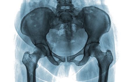
X-ray:
X-rays are suitable for detecting bone fractures, bone infections, joint sprains, joint wear and spinal deformities, and can also be used to examine the lungs, certain soft tissues and the abdominal organs. The recording can be made:
- about the bones that make up the body's skeletal system due to injury, degenerative deviation, suspected fracture, bone change, exclusion of other pathological processes, developmental disorder;
- bruises from joints, chronic pain, etc. in case of;
- about complaints that are a combination of certain chest and abdominal symptoms, such as acute abdomen, PTX-HTX, TBC, etc.,
- otorhinolaryngological changes, inflammatory changes of the sinuses, as well as
- before surgeries and
- as a supplement to a medical examination.
X-ray examinations do not involve any preparation and are absolutely painless. It is important that the patient receiving the X-ray cannot wear jewelry, glasses, or metal objects during the examination. During the examination, a picture is taken of the affected body part in a motionless position, from one or more directions.
Bone density measurement:
The bone densitometry is used to measure the mineral content of the bone and to map the condition of the bones . It can be used to determine the risk of osteoporosis and fracture and to monitor the success of the treatment.
In 70-80% of cases, the cause of osteoporosis is genetic, in addition, lack of physical activity, smoking, excessive alcohol consumption, a diet low in calcium and vitamins, low vitamin D intake, and the existence of certain diseases (e.g. hormonal diseases, diseases of the intestinal system and malabsorption, abnormal thinness, anorexia, chronic kidney and liver failure, thyroid hormone overproduction, asthma) and the long-term use of certain drugs (e.g. steroids, drugs that weaken the immune system, epilepsy drugs).
The bone density test is primarily recommended for women over 50 years of age, and for those patients who have had osteoporosis in their family before. During the 15-20 minute examination (before which all jewelry must be removed), the lumbar spine, hips on both sides and, in some cases, the entire body will be examined in the supine position.
Ultrasound examinations:
Ultrasound is a painless examination, with the help of which many problems and organ changes can be discovered. Its use is justified for many problems affecting our body, but it is most common for gastroenterological, gynecological, internal medicine, endocrinological, urological, vascular (Doppler ultrasound) and joint complaints.
The following ultrasound examinations are available at the Duna Medical Center:
Abdominal/pelvic ultrasound : creates an image of every organ from the diaphragm to the pubic bone, so we get more detailed information about the abdominal organs, soft tissues (liver, gall bladder, bile ducts, pancreas, spleen, kidneys, bladder) and lymph nodes . This examination is most often recommended in the case of abdominal complaints of unclear origin, urination and menstrual problems. In addition to these, it is also suitable for monitoring known diseases and control tests for cancerous changes .
Ultrasound examination of the soft tissue of the neck, thyroid gland, blood vessels, testicles, joints: the use of ultrasound is justified in the case of neck pain, swallowing discomfort, thyroid gland dysfunction, or for the purpose of checking known diseases, neoplastic diseases, post-operative control examinations or screening tests.
The pathological condition or damage of blood vessels (arteries, veins, aorta) can also be checked with the Doppler ultrasound procedure.
Examination of the joints and the testicle may be necessary in case of muscle injuries, changes and inflammations.
Cardiac ultrasound examination: this examination takes place in the presence of a specialist, during which a moving image of the heart is obtained, so that the heart cavities, the walls that border them, the valves and the pericardium can also be examined. A gentle and painless examination with no harmful effects. It can be used to detect, for example, post-infarction wall motion disorders, scarring, valvular diseases, abnormal blood flow between the chambers of the heart, fluid accumulated in the pericardium or blood clots formed in the chambers of the heart.
Performing an echocardiogram is definitely justified after the first cardiology consultation, as it greatly helps the cardiologist to accurately assess the condition of the heart.
Pregnancy ultrasound: in our institution, within the framework of pregnancy care, we continuously perform pregnancy 2D, 3D, 4D ultrasound examinations from the early stages of pregnancy, which help to establish pregnancy and monitor the development of the baby.