
 Mammography is warranted in cases of palpable lumps in the breast and other complaints, and regular screening is recommended for those over 40 years of age. The examination does not require serious preparation, but the patient needs to bring previous findings, their images, and possible biopsy results.
At Duna Medical Center, all breast screening and diagnostic procedures are available, including the most important breast interventions: aspiration cytology, core biopsy, stereotaxic vacuum assisted breast biopsy. Vacuum assisted removal of certain marginal lesions and tumour masses (papillary, B3 lesions) is performed without anaesthesia on an outpatient basis.
It is possible to request a second opinion at our institution. In the case of breast lesions not detected elsewhere, stagnation of the examination or unsuccessful, unsuccessful or very complicated breast biopsy, it is possible to have a personal consultation, additional tests, sampling. For this purpose, the patient must bring the images and findings from a previous visit to another institution on CD or paper.
The radiology department of Duna Medical Center is equipped with modern, state-of-the-art equipment. In addition to the quality of the equipment, the expertise and experience of the radiologist and the radiology assistants have a decisive influence on the evaluation of the examinations. As we are an accredited training centre of Semmelweis University, we also train radiologists preparing for the breast licensing examination. During mammography examinations, our patients are examined by breast radiologists with such specialist examinations and many years of experience.
In the hands of our skilled, experienced, patient and empathetic mammography assistants, ladies are safe. Feel free to come in for an exam even if you are about to have your first mammogram or have had a negative mammogram experience elsewhere.
We are proud that our national mammography assistant textbook was written by the team at Duna Medical Center.
Both the American and Dutch breast screening programs are based on the Hologen - American mammography equipment we use. For detailed information about Duna Medical Center's state-of-the-art radiology equipment, click here: https://dunamedicalcenter.org/hirek/csucstechnologia-a-duna-medical-centerben
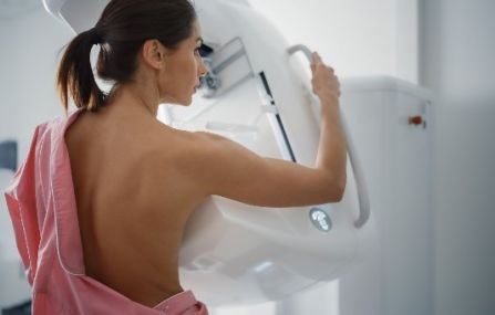
During the mammography examination, the standard mammography images (4 images) of the two breasts are always taken together with 2D and 3D tomosynthesis. The device also produces synthetic 2D images, which helps to detect tumours.
If there is an area in the breast that needs to be cleared and the radiologist considers it necessary, additional mammograms can be taken, which are free of charge (included in the price of the mammogram). There is no call-back or waiting time at our institution, and the additional breast ultrasound scan can be performed immediately.
Attention! Absolute contraindication for X-ray examinations is pregnancy or suspicion of pregnancy. A written negative declaration of pregnancy is required. If the patient is uncertain whether she is pregnant, the examination cannot be performed. The radiology assistant may not take the scan until he/she is satisfied that he/she has sufficient information about a possible pregnancy. Of course, mammography assistants are particularly careful about radiation exposure.
Mammography is recommended as a screening test for women without complaints, as well as in cases where previously non-palpable lumps, secretions, nipple indentation, signs of inflammation, pain appeared in the breast.
In accordance with international recommendations, regular mammography screening is recommended for everyone over the age of 40. The earlier a breast examination is performed and breast cancer is recognized, the more effective the treatment can be. That is why it is extremely important for women to undergo regular screening.
At a younger age, the radiologist decides on the need for this - depending on age and risk factors - and based on the complaints.
At our hospital , mammography examination can be done without a referral, at a time agreed in advance by phone or online.
The test consists of 4 basic recordings, which takes about 10 minutes.
The image is taken in a standing position (depending on the condition, rarely sitting) with special X-ray equipment (in our clinic, with 2D, 3D tomosynthesis, i.e. making layered images).
In order to ensure adequate image quality, we apply the necessary pressure on the breasts, which, apart from mild discomfort, cannot be said to be painful. The recordings are reviewed by the radiologist, followed by a physical examination and an ultrasound examination of the breasts and both armpits.
In certain, not clearly negative cases, it may become necessary to take additional special mammographic images, which depict the given area more precisely and in more detail.
In the case of sensitive breasts, it is advisable to schedule the mammography examination for the first two weeks after the first day of menstruation, but any deviation from this does not affect the evaluation, quality, and accuracy of the results. Adaptation to menstruation should only be considered based on the above comfort aspects.
Before the examination, it is advisable to remove cosmetics, creams, and antiperspirants applied to the skin.
Mammography imaging is performed with low-energy ionizing radiation, so the radiation risk is negligible. Mammography has no side effects.
Our X-ray assistants take special Eklund images of the implanted breast. With this technique, the implants are not put under pressure, only the examination of the breast tissue and the armpit region is possible. The examination does not involve more discomfort than traditional mammography.
The summary mammographic findings are handed over by the radiologist performing the examination immediately after the joint evaluation of the completed mammographic images and the US examination.
Mammography images can also be requested in digital form on a CD, which we ask you to notify in advance. However, these recording series cannot be viewed on a home PC, they can be opened in the image viewer of a high-resolution computer workstation specifically suitable for medical use.
Vacuum-assisted excision (ambulatory removal of certain /B3/ breast lesions using vacuum)
There are breast lesions that are not malignant tumors (not breast cancer), but they are also not completely benign processes, which is why they must be completely removed from the breast.
The reason for this is that these borderline (so-called "B3") deviations are not necessarily the same throughout their entire area, so there may be a hidden malignant part in them, or if they are not removed, they may later develop into a tumor.
These changes are most often papillary lesions (papillomas) , but several similar processes are also included ( radial scar , atypical ductal hyperplasia, i.e. ADH, AIDEP, FEA, mucocele-like lesions, classic lobular neoplasia, etc.)
Vacuum-assisted excision is the most modern so-called "minimally invasive" procedure.
Unlike surgery, there is no need for anesthesia, no hospitalization, and no scar line on the breast.
Procedure of the intervention:
The intervention is carried out with ultrasound or mammographic guidance, in a supine or side-lying position, under local anesthesia (without anesthesia), the duration of which is approx. 20 minutes. The preliminary preparation, aiming and dressing total approx. it takes an hour. There is no need to prepare for pain, the patient may feel minor discomfort at most.
The intervention is performed on an outpatient basis, after which the patient can leave. The histological findings will be completed within 5-10 working days, the results will be handed over personally by the doctor who performed the intervention, and he will explain any further actions that may need to be taken.

If you require a medical report in a foreign language (English, German) please indicate this to our staff during your visit, as preparing these involves an additional cost. The documents are prepared within 3-10 working days.

-
From 33 000 Ft
-
Complex breast examination, breast screening: digital low-dose mammography with 3-dimensional tomosynthesis + breast and axillary ultrasound examination with elastography in medically justified cases + medical physical examinationFrom 66 000 Ft
-
Stereotaxic breast biopsy, 3-dimensional, vacuum-assisted (includes histological processing)From 454 000 Ft