
The Leading Private Hospital in Budapest Continues as an Independent Brand with Renewed Strength
A new chapter begins in the history of one of Budapest’s most advanced private healthcare institutions. Building on the experience gained through its previous international partnership, the hospital is once again operating under the name Duna Medical Center as of today, ...

Birth Statistics 2025
For Duna Medical Center, obstetrics is not merely a service, but a responsibility and a calling. The 2025 obstetrics data clearly reflect a professional approach that prioritizes safety, medical expertise, and the individual needs of expectant mothers alike.

Vaccinations Recommended and Safe During Pregnancy – 2025 Guidelines
We have compiled a list of vaccines that can be administered during pregnancy. It is important to consult your doctor before receiving any vaccine.
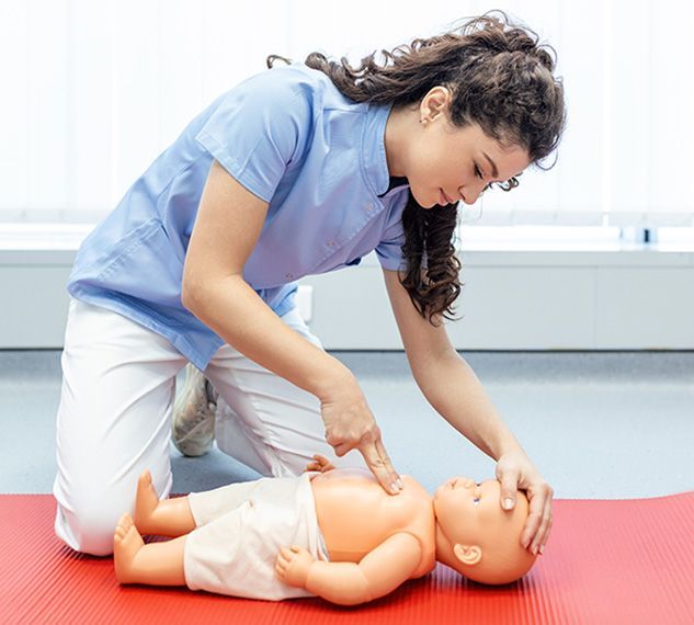
Baby First Aid Course
Duna Medical Center is launching a unique programme, an infant first aid course, to help parents during one of the most important times of parenthood.
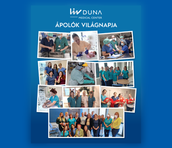
Celebrating International Nurses Day
Let us take this day to express our deepest gratitude to all nurses for their selfless service and commitment to care.

Stipendium Hungaricum
Do you need help in submitting the medical certificate for Stipendium Hungaricum scholarship program?


How to put together our meals correctly?
The cornerstone of a healthy diet is a varied, regular and balanced meal rich in nutrients. By regularity, we mean at least 3 main meals a day consumed at approximately the same intervals, in addition to which it may be worthwhile to include 1 or 2 snacks according to individual ...

Influenza is a quick fix
Vaccination makes you sick, but it gives you lifelong immunity and you can only catch the flu once a season - these are the most common misconceptions about the flu. Read our blog to find out why you should take flu season seriously every year!


We will rid you of hemorrhoids with a gentle procedure!
There is a procedure that allows us to get rid of our hemorrhoid complaints without cutting, and thus almost painlessly - no matter how serious they are. We asked Dr. Péter Sipos, surgeon of the Duna Medical Center.

The hemorrhoids no one likes to talk about
Hemorrhoids are a very common disease, affecting 50% of the adult population, yet we don't like to talk about it. However, it would be important to know what types there are and when we should consult a specialist in order to prevent an unpleasant intervention. Dr. Péter ...
Nose and throat tonsil removal gently
New at the Duna Medical Center private hospital! We are introducing a new, modern, innovative procedure for the removal of the throat and nasopharynx: with the use of BiZact™ tonsillectomy scissors, the procedure is carried out in a gentle, bloodless manner, which has ...